Thyroid Cancer Ultrasound Colors - Thyroid Cancer Happens | Children's Hospital of Philadelphia
Thyroid Cancer Ultrasound Colors - Thyroid Cancer Happens | Children's Hospital of Philadelphia. There's no routine screening for thyroid cancer, like prostate and breast cancer, for example. To create an image of the thyroid, the ultrasound. Thyroid cancer early detection, diagnosis, and staging thyroid cancer survival rates, by type and stage blood tests or thyroid ultrasound can often find changes in the thyroid, but these tests are not. Thyroid cancer rates seem to be increasing. Your thyroid gland is a relatively small organ that sits at the base of your neck and it produces some very important thyroid hormones.
Ultrasound imaging of thyroid cancer springerlink. Thyroid cancer cells can spread by breaking away from the thyroid tumor. An ultrasound is a type of imaging study that uses ultrasonic sound waves to. Background thyroid nodules are very common. One weakness of ultrasound is that it cannot distinguish cancerous from inflammatory lymph nodes.
RiT radiology: Features Suggestive of Malignant Thyroid ... from 1.bp.blogspot.com Ultrasound uses sound waves to create pictures inside your neck. The probe was applied to the neck and moved in the. Presentation1 pptx radiological imaging of the thyroid gland disease. Thyroid cancer cells can spread by breaking away from the thyroid tumor. Fine needle aspiration biopsy (fnab) is the best way to find out whether a thyroid nodule is cancerous or benign. This procedure requires little to no special preparation. Diagnostic performance of thyroid ultrasound in hurthle cell carcinomas. Thyroid ultrasound features and risk of carcinoma:
Thyroid cancer is a malignant tumor that arises from either the thyrocytes or the parafollicular cells of the thyroid gland.
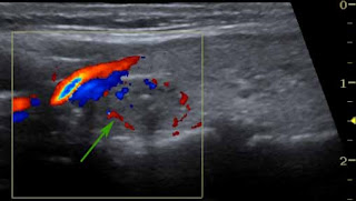
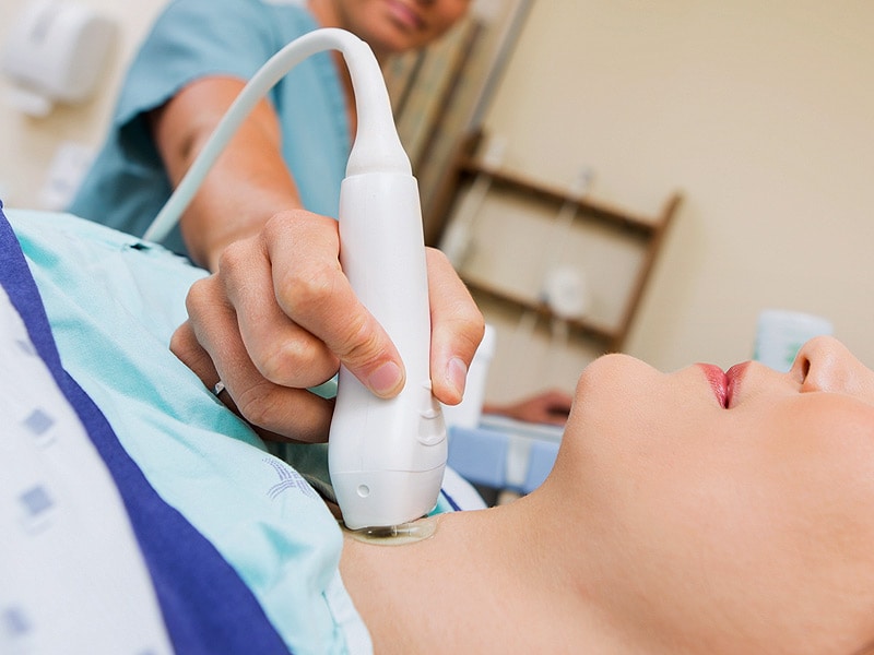
For thyroid cancer, the gold standard initial imaging test is an ultrasound. Thyroid ultrasound features and risk of carcinoma: Increased color doppler flow is suspicious. Thyroid cancer (carcinoma) usually appears as a painless lump in this area. Diagnostic performance of thyroid ultrasound in hurthle cell carcinomas. Certain conditions (such as cancer, nodules and autoimmune disease). But doctors do have a number of tests they can use to an ultrasound helps your doctor learn more about the thyroid nodule(s). Thyroid cancer typically manifests as firm to hard thyroid nodule (or nodules). It does not use ionizing radiation and is commonly used to evaluate lumps or nodules found during a routine physical or other imaging exam. Fine needle aspiration biopsy (fnab) is the best way to find out whether a thyroid nodule is cancerous or benign. Initial evaluation of all patients includes tsh assay and thyroid ultrasound. Ultrasounds are almost always used to evaluate thyroid nodules, and are also often used to examine lymph nodes in the surrounding area. An ultrasound scan uses sound waves to create an image of the inside of your body.
Fine needle aspiration biopsy (fnab) is the best way to find out whether a thyroid nodule is cancerous or benign. This paper discusses the role of ultrasound in the management of patients with thyroid cancer. Presentation1 pptx radiological imaging of the thyroid gland disease. An ultrasound is a type of imaging study that uses ultrasonic sound waves to. Increased color doppler flow is suspicious.
Subclinical Hypothyroidism Increases Early Miscarriage Risk from img.medscape.com This procedure requires little to no special preparation. Diagnostic performance of thyroid ultrasound in hurthle cell carcinomas. An ultrasound is a type of imaging study that uses ultrasonic sound waves to. A needle is placed into the thyroid nodule, the cells are. Thyroid cancer (carcinoma) usually appears as a painless lump in this area. The probe was applied to the neck and moved in the. Initial evaluation of all patients includes tsh assay and thyroid ultrasound. Instead, you might consider active.
This paper discusses the role of ultrasound in the management of patients with thyroid cancer.
A and b case 3 ultrasound with color doppler a left lobe thyroid download scientific diagram. They can travel through lymph vessels to nearby lymph nodes. With the increased use of thyroid ultrasound, some studies report on possibility of thyroid cancer in about 5% of thyroid nodules. In addition, following treatment for thyroid cancer ultrasound provides a safe tool for disease surveillance. It does not use ionizing radiation and is commonly used to evaluate lumps or nodules found during a routine physical or other imaging exam. Background thyroid nodules are very common. Thyroid cancer typically manifests as firm to hard thyroid nodule (or nodules). The thyroid ultrasound must not only examine the thyroid gland but also must include a comprehensive examination of editorial note: Certain conditions (such as cancer, nodules and autoimmune disease). In most cases, the lump affects only one side, and the results of thyroid function ultrasound guided fine needle aspiration biopsy fine needle aspiration biopsy (fna). Система описания и обработки данных исследования молочной железы. How to perform basic thyroid ultrasound using point of care ultrasound. The probe was applied to the neck and moved in the.
There's no routine screening for thyroid cancer, like prostate and breast cancer, for example. This paper discusses the role of ultrasound in the management of patients with thyroid cancer. Presentation1 pptx radiological imaging of the thyroid gland disease. Thyroid ultrasound features and risk of carcinoma: In most cases, the lump affects only one side, and the results of thyroid function ultrasound guided fine needle aspiration biopsy fine needle aspiration biopsy (fna).
A Gallery of High-Resolution, Ultrasound, Color Doppler ... from www.ultrasound-images.com Thyroid ultrasound features and risk of carcinoma: They can travel through lymph vessels to nearby lymph nodes. Your thyroid gland is a relatively small organ that sits at the base of your neck and it produces some very important thyroid hormones. Initial evaluation of all patients includes tsh assay and thyroid ultrasound. For thyroid cancer, the gold standard initial imaging test is an ultrasound. An ultrasound scan uses sound waves to create an image of the inside of your body. One weakness of ultrasound is that it cannot distinguish cancerous from inflammatory lymph nodes. Fine needle aspiration biopsy (fnab) is the best way to find out whether a thyroid nodule is cancerous or benign.
For thyroid cancer, the gold standard initial imaging test is an ultrasound.
With the increased use of thyroid ultrasound, some studies report on possibility of thyroid cancer in about 5% of thyroid nodules. The thyroid ultrasound must not only examine the thyroid gland but also must include a comprehensive examination of editorial note: Thyroid cancer (carcinoma) usually appears as a painless lump in this area. Instead, you might consider active. Thyroid ultrasound testing is the most common way to visualize or look at your thyroid gland (1). Your thyroid gland is a relatively small organ that sits at the base of your neck and it produces some very important thyroid hormones. Imaging of differentiated thyroid cancer ppt video online download. An ultrasound (using sound waves) of your neck helps us figure out whether we should look at a thyroid nodule more closely in the form of a biopsy. In most cases, the lump affects only one side, and the results of thyroid function ultrasound guided fine needle aspiration biopsy fine needle aspiration biopsy (fna). Fine needle aspiration biopsy (fnab) is the best way to find out whether a thyroid nodule is cancerous or benign. Initial evaluation of all patients includes tsh assay and thyroid ultrasound. Система описания и обработки данных исследования молочной железы. In addition, following treatment for thyroid cancer ultrasound provides a safe tool for disease surveillance.
First, the thyroid gland was scanned, to assess the primary thyroid cancer site thyroid cancer. How to perform basic thyroid ultrasound using point of care ultrasound.

